Electronic english version since 2022 |
The newspaper was founded in November 1957
| |
Science - to practice
How physicists have made breakthroughs
in medical diagnostic methods
Today, medicine widely uses the knowledge accumulated over hundreds of years in physics. There are many physicists in the world that develop methods and technologies for medicine. Others work in healthcare institutions, handling complex equipment: particle accelerators, CT, MRI, PET facilities, others. They are called medical physicists and the field of science in which they work is medical physics. This is a section of applied physics that uses physical principles, methods and techniques in practice and research for the prevention, diagnosis and treatment of diseases in order to improve human health and well-being. We talked about the origin and development of these methods, their diversity nowadays to DLNP researcher Vladislav ROZHKOV.
The discovery of X-rays. The beginning of a new era
 Physics and medicine have been close to each other in their development. And scientists almost immediately began to apply a large number of discoveries in the field of physics in medicine. At the turn of the 19th and 20th centuries, a revolution occurred in science: scientists penetrated inside the atom and later, inside the nucleus. In the twentieth century, physicists and doctors made a number of outstanding achievements.
"Back in 1887, Nikola Tesla experimented with various types of radiation," Vladislav Rozhkov noted. "He recorded visible light, ultraviolet radiation and special rays that could penetrate objects. And only with the discovery of X-rays by Wilhelm Conrad Roentgen in 1895, Tesla understood what he dealt with. Subsequently, he even improved the X-ray tube and wrote several scientific publications on the nature of this radiation. Literally a few months after Roentgen had published his scientific paper, X-ray machines were widely used in medicine."
Thus, since the beginning of the century, X-rays, for the discovery of which Roentgen was awarded the Nobel Prize in Physics in 1901, have become an integral part of any medical institution, allowing one to look inside a person, literally "to see" his bones. It was the beginning of a new era. Military surgeons were especially interested in X-rays and Russia did not lag behind Europe in this regard.
"War Minister Petr Vannovsky even ordered the allocation of 5 thousand rubles, for which the Military Medical Academy purchased two X-ray machines and consumables for them in Germany - photographic plates and chemicals. In 1897, Russian doctors successfully used X-rays in the Greco-Turkish War. Thanks to Alexander Popov, X-ray machines also appeared in the Navy. The cruiser Aurora had an X-ray room where assistance was provided to sailors that suffered from bullet and shrapnel wounds," the scientist said.
Radiotherapy and radioactivity
Chicago medical student Emil Grubbe is the one that made people think that X-rays affect biological objects. It was with him that radiotherapy began: Grubbe developed a scheme that has not changed to this day and is used in all X-ray therapy rooms, when the patient with the source is in one room and the "screen" is in another, where the doctor does not receive a dose. On 27 January, 1896, the idea of radiotherapy was born and two days later, the first radiotherapy session was carried out.
At the turn of the 19th and 20th centuries, the concept of radioactivity also appeared. French physicist Henri Becquerel noticed that uranium salts that he studied with the Curies, leave traces on the skin. There is a story that once, Henri leaving the laboratory, took a piece of radioactive ore with him and put it in his breast pocket, then forgot about it, walked with it for about a week. Ulcers appeared on the chest in this place and then the scientist made certain conclusions. His discovery served as a starting point for the development of nuclear physics and subsequently nuclear medicine and radiation therapy.
The occurrence of various side effects from the action of radiation in specialists working with it prompted them to think about the need for protection. Doctors understood the danger of radiation and began to wear special rubberized lead spacesuits.
"Despite it, miraculous properties were attributed to radium for many years and its radioactivity was "closed to" Vladislav Rozhkov says. "Some businessmen of that time decided to use it to get rich. For example, they would put a small piece of radium in a container with water and say that the water would be purified and filled with radiant energy. Such revigators - water filters were sold almost until the 1960s. At first, the human body really did get a boost of energy from such a drink, for all its strength and resources were directed at removing the radioactive substance from the body." Water, sugar and coffee were also X-rayed. It was believed that it made the products healthier. However, in reality, X-rays only disinfected them. Irradiated golf balls were also sold. It was believed that the owner of such balls would score nine times out of ten.
Development of accelerators and the production of isotopes. The birth of nuclear medicine
On 27 February, 1932, the English experimental physicist James Chadwick discovered the neutron, an elementary particle that has no electric charge. For his discovery, Chadwick was awarded the Nobel Prize in Physics in 1935. The neutron was the last of the three fundamental particles that make up an atom (electron, proton, neutron). His discovery indicated the occurrence of a new type of force in nature - nuclear.
In the 1930s, ideas were born and there were many physical developments that later turned into nuclear-physical technologies in medicine. In complex physical facilities - particle accelerators - beams of electrons, protons and high-energy photons are obtained for the treatment of malignant neoplasms. In the late 1920s - early 1930s, the first charged particle accelerators were built.
In 1928, the first linear accelerator was patented. Linear accelerators allow achieving high speeds of light charged particles (primarily electrons). They can be used to irradiate matter, produce isotopes and test materials for radio resistance. In medicine, linear accelerators are widely used as the main element (source of X-ray, electron, proton radiation) of devices for radiotherapy and radiosurgery. Achievements in accelerator physics have created the foundation for a wider use of radioactive isotopes. In the early thirties, Ernest Lawrence built a resonant ring accelerator - a cyclotron. It was on cyclotrons that most artificial radioactive isotopes were discovered that found application in nuclear medicine and radiation therapy. The time of the beginning of isotope deliveries that dates back to 1946, is considered the date of the birth of today's nuclear medicine that uses radioactive radiation of isotopes for diagnostics and therapy. "In Russian practice, the possibility of carrying out research with radioactive isotopes was ensured by the commissioning of the first cyclotrons in 1937, 1944, 1947 and the first nuclear reactor in the country in 1946. Since 1948, regular production of radioactive isotopes for scientific and medical purposes has been carried out: sodium, potassium, phosphorus, iodine, hydrogen, chromium, iron and others," the scientist emphasized.
Radioisotope diagnostic methods: gamma camera, SPECT, PET
In addition, methods for obtaining images of human organs using radiopharmaceuticals were developed in diagnostics. In 1958, Hall Anger developed a gamma camera - a device for obtaining a two-dimensional image of the distribution of gamma sources in the object being studied. The occurrence of collimators in the structure of the gamma camera allows using only gamma quanta of the selected area for image reconstruction that in turn helps to determine the position of the radiation source in space. Today, the method is widely used in investigations of the thyroid, pancreas and small laboratory animals.
The development of these diagnostic methods using computer technology in real time became a basis for single-photon emission computed tomography (SPECT). In SPECT, a radionuclide emitting gamma quanta is used to obtain an image. The radionuclide is part of the radiopharmaceutical that accumulates in various organs and tissues of the patient. SPECT today is a system consisting of several gamma cameras rotating around the patient and a table moving in a horizontal plane. Today, SPECT is one of the best radioisotope research methods and is most often used for targeted analysis of a particular organ.
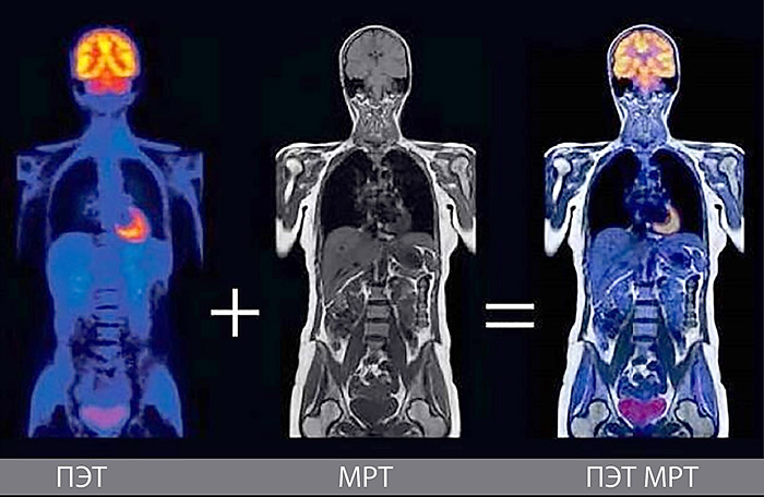
Positron emission tomography (PET), like SPECT, is a method of radioisotope diagnostics that allows obtaining information about the functioning of a selected organ or the entire body. However, PET uses isotopes that emit positrons, rather than gamma quanta - elementary particles equal in mass to an electron and positively charged.
PET consists of a fixed ring of detectors and a movable table on which the patient is placed. The radioisotope in the radiopharmaceutical is administered to the patient intravenously, after which it circulates in the blood and reaches the organ being examined. When the emitted positron meets an electron in the environment in which it is located, annihilation occurs, that is, the particles turn into two gamma quanta flying off in opposite directions. Since they reach the detectors simultaneously, it is possible to determine the line on which the annihilation occurred. Many of these lines allow identifying where the radioisotope accumulates. "This is an excellent method for diagnosing oncological diseases at early stages," Vladislav Rozhkov notes. "The patient is injected with a radiopharmaceutical based on a glucose molecule, in which one of the atoms is replaced by radioactive fluorine. In addition to the brain that widely consumes glucose, high consumption of this molecule is also characteristic of tumor cells. And the occurrence of oncological disease can be judged by the places where the drug accumulates."
CT and MRI - a breakthrough in diagnostic and visualization methods
The first CT scanner was designed in 1969 by English engineer and physicist Godfrey Hounsfield that worked at the Amy Records Studio, where most of the Beatles' albums were recorded. He proposed developing a device that could create three-dimensional images of the brain using X-rays and the company approved the project. Hounsfield realized that multiple projections connected to each other can form a three-dimensional image. And it allows one to accurately determine the localization and prevalence of the pathological process.
Early prototypes were tested on the human brain - the first patient was diagnosed with a brain tumor that was then successfully removed. The scan results revolutionized medical imaging and diagnostic procedures. The new method quickly replaced painful, dangerous and sometimes useless procedures. The joint work of Hounsfield and mathematician from South Africa Allan Cormack was awarded the Nobel Prize in 1979.
In 1944, the phenomenon of electron paramagnetic resonance was discovered, in 1946 - the phenomenon of nuclear magnetic resonance, and in 1973 - the magnetic resonance tomograph was developed.
"The essence of this phenomenon is that the nuclei of some atoms, when placed in a magnetic field, are capable of absorbing energy in the radio frequency range and emitting it after the effect of the radio frequency pulse ceases. In this case, the intensity of the constant magnetic field and the frequency of the radiofrequency magnetic field must strictly correspond to each other that is called nuclear magnetic resonance," Vladislav Rozhkov explains.
A magnet produces a magnetic field in the tomograph. It is made three-dimensional using gradient coils, inside which there is a radiofrequency coil that emits and receives radiofrequency pulses from the nuclei of the object being examined. Modern MRIs allow you to get a three-dimensional "map" of the distribution of hydrogen nuclei present in the human body, they display soft tissues well, unlike CT that visualizes bone structures better.

"In modern medicine, combined methods are increasingly used, such as SPECT/CT, PET/CT, CT/MRI and other combinations: one image is superimposed on another and a more complete picture is obtained. Such systems are called multimodal. They are very difficult to modernize and make better, the scientist admits. However, science moves towards improving detectors that in the foreseeable future will be able to register not only the interaction coordinates, but also the registered energy of this radiation. In addition, the methods and algorithms for reconstructing and analyzing the obtained images can change quite significantly. Machine learning is of great importance here," he notes.
Kseniya MORUNOVA